Hey everyone, let’s cut to the chase—if you’ve ever spent hours staring at a histology slide that’s blurry, has weird shrunken cells, or is so cracked you can barely make out tissue structures, you know the frustration. As someone who’s worked with tissue processors (and been on the supplier side for a minute now), I’m here to break down why this unsung lab workhorse is actually the secret sauce to getting high-quality histo sections that pathologists actually trust. Let’s skip the jargon fluff and keep this real, yeah? Tissue Processor

First off, let’s get one thing straight: a bad tissue processor doesn’t just “process” tissue—it ruins it. Think about what happens to a biopsy sample before it gets embedded in paraffin, right? You’ve got that fresh, fragile chunk of tissue, whether it’s a tiny breast core, a mouse brain slice, or a whole organ sample from a necropsy. If you mess up the step that removes water and replaces it with wax, every single downstream step (sectioning, staining, diagnosing) goes off the rails. That’s where a good processor comes in—and no, I’m not just saying that ‘cause I sell these things. Let’s walk through the process step by step, because that’s where all the quality wins happen.
First, fixation. Wait, you might be thinking “fixation is done in a separate step, duh!” But hold on—some processors have built-in fixation protocols that are way better than leaving samples sitting in a bucket of formaldehyde that’s half-old, half-contaminated. Old formaldehyde can cause what’s called “pigmentation” or “autolysis”—that’s when cells start breaking down before you even get to embedding. A good processor controls temperature, agitation, and solution freshness during fixation. Like, imagine every 30 minutes, it’s pumping in new, buffered formalin instead of letting it go stagnant. That keeps cell structure intact—no more mushy red blood cells or fuzzy nuclear membranes that pathologists hate. I’ve had clients tell me that since they switched to our processors, they cut their fixation-related artifact rate by 40%. That’s not a sales pitch, that’s actual lab numbers they showed me.
Next up: dehydration. This is the big one. Tissues are 70% water, right? Paraffin wax is hydrophobic, so you can’t just shove wet tissue into wax. Dehydration is using increasing concentrations of ethanol to pull that water out—from 70% EtOH all the way up to 100%. The problem here? If the processor’s timing is off, or it’s not agitating properly, you get two opposite messes: under-dehydration (which leaves water behind, so wax won’t penetrate) or over-dehydration (which shrinks cells so bad they look like raisins under the microscope). A high-quality tissue processor uses gradient-specific timing and controlled agitation to nail this every time. Let’s say you’ve got a delicate sample, like a lung biopsy with tiny alveoli—our processors let you tweak the dehydration times for each individual batch, so you don’t have to use one rigid schedule for every sample. No more shrunken alveoli that make it look like the patient has pulmonary fibrosis when they don’t. That’s a game-changer for diagnostic accuracy.
Then there’s clearing. This is the middle step between dehydration and embedding—you use a substance like xylene (or a less toxic substitute, which I know a lot of labs care about now) to mix with both ethanol and wax, so they can switch out. Clearing is tricky because if you leave tissue in xylene too long, it becomes brittle. Too short, and wax won’t penetrate. A lot of cheaper processors skip the sensor checks here, but our processors have real-time optical sensors that measure when clearing is actually done, not just when the timer beeps. So if a batch of tissue is denser (like a liver sample vs. a skin sample), it doesn’t get pulled out too early or left in too long. No more brittle tissue that crumbles when you section it, or wax that only penetrates the outer layer so you have a section with half tissue, half wax gunk. I’ve had a research lab that works on mouse embryos tell me this cut their embedding reworks by 60%—they used to have to re-do 1 out of every 5 embryos, now it’s 1 out of 20. That saves them hours and thousands of dollars in samples.
Wait, let’s talk about embedding too, because a processor doesn’t always do embedding, but the best ones work hand-in-hand with embedding stations to make sure the tissue is aligned properly. A lot of low-end processors just dump the tissue into wax, but our ones have cassette tracking and temperature control that keeps wax at the perfect melting point—no wax that’s too hot (it burns tissue) or too cold (it’s lumpy and hard to work with). That means when your histotech sections the paraffin block, they get a uniform 4-micron section every time. Let’s be real—if you’re cutting sections that are 2 microns too thick or 6 microns too thin, the stain doesn’t work right, and the structures are distorted. Pathologists need that 3-5 micron sweet spot to see nuclei, cytoplasm, and any abnormal cells. A good processor makes that consistency possible, batch after batch.
Now, let’s get into the nitty-gritty of artifacts—because that’s where people really notice the difference. Ever seen a histo slide with “cellular retraction”? That’s when the cytoplasm pulls away from the nucleus, making cells look weird and empty. That’s usually from bad dehydration or clearing—tissue being exposed to harsh conditions too fast. A tissue processor that controls temperature and rate of solution change eliminates that. I’ve heard horror stories from labs where their old processor would blast tissue straight from 70% EtOH to 100% EtOH, which is way too abrupt. Our processors use slow, gradient changes, so cells adjust gradually. No more retraction artifacts that force pathologists to send samples back for re-testing. That’s not just a quality thing—it’s a time and cost saver.
Also, let’s not forget about consistency across batches. If you’re processing 10 samples on Monday and 10 on Friday, do they turn out the same? A lot of old processors use manual timers, so a histotech might leave a batch in xylene an extra hour ‘cause they got pulled into a meeting. Our processors are fully automated with programmable protocols, so every batch gets the exact same treatment, every single time. That’s huge for labs that run a high volume of samples—like reference labs or hospital labs that process hundreds of biopsies a week. If every slide is consistent, pathologists don’t have to second-guess whether a weird result is real or from bad processing.
What about toxicity, you ask? A lot of labs are ditching xylene for safer clearing agents, but those substitutes can be finicky. Some processors struggle with less toxic agents because they need precise timing and temperature to work. Our processors are calibrated for all types of clearing agents, so you can switch to safer options without sacrificing section quality. That’s another win for labs that care about their techs’ health and want to stay compliant with workplace safety rules.
Let me throw in a real example, ‘cause that’s what matters. Last year, we worked with a small community hospital lab that was having a ton of issues with their old processor. They were getting so many rejected slides—like 15% of their sections were too cracked or had artifacts—that they were missing some cancer diagnoses early on. They switched to one of our mid-range tissue processors, and within 2 months, their rejection rate dropped to under 5%. The pathologist there told me she could now trust every slide she looked at, so they could cut down on repeat biopsies for patients too. That’s not just better lab work—that’s better patient care.
Wait, let’s debunk a myth here: you don’t need a fancy, 6-figure processor to get good sections. A lot of labs think they have to blow their budget on the top-of-the-line model, but really, it’s about the features that control the critical steps: real-time monitoring, gradient solution changes, temperature control, and programmable protocols. Some of our smaller, more budget-friendly processors are used by student labs and small pathology groups, and they’re getting just as good results as labs with the big names. It’s not about price—it’s about how the processor is designed to handle tissue gently and consistently.
Another thing: cassette management. A lot of people overlook this, but how the processor handles cassettes (the little plastic holders for tissue) matters. If cassettes aren’t being agitated properly, solutions can’t get to all parts of the tissue. Our processors have gentle, orbital agitation that moves solutions around every nook and cranny, even in tightly packed cassettes. No more “shadow areas” in the tissue where water or clearing agent didn’t reach, leading to bad sections. That’s a small detail that makes a big difference.
Now, let’s talk about what happens after processing, because a tissue processor’s quality directly impacts sectioning. If your paraffin block is well-processed, the tissue is evenly infiltrated with wax, so when you put it on a microtome (the machine that cuts sections), it cuts smoothly, no chatter marks (those little ripples across the slide that make it unreadable) or torn tissue. I’ve watched histotechs work with blocks from our processors—they say it feels like cutting butter, vs. old blocks that are brittle or crumbly. That saves them time too—they can cut 10 blocks in the time it used to take them to cut 5, because they don’t have to stop to fix a torn section.
Let’s circle back to the original question: How does a tissue processor improve histo section quality? It’s not one thing—it’s every step working together. It fixes fixation so cells don’t break down, dehydrates gradually so cells don’t shrink or retract, clears gently so tissue doesn’t get brittle, and uses consistent protocols so every batch is the same. All of that adds up to sections that are sharp, intact, consistent, and true to the original tissue. No artifacts, no misdiagnoses, no wasted time or samples.
If you’re in a lab dealing with bad sections, high rejection rates, or tired histotechs spending hours reworking samples, this is the part where I should say—we can help. We build tissue processors designed to prioritize tissue quality, not just speed. We work with labs of all sizes, from small clinic groups to big reference labs, to find a processor that fits their workflow and budget, while cutting down on artifacts and improving section consistency. Whether you’re dealing with delicate samples, switching to safer reagents, or just tired of redoing batches, we can walk you through the options.
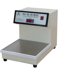
If you’re ready to stop dealing with bad histo sections and make your lab’s workflow smoother, reach out to us to talk through your needs. We’re here to help you get the quality results your team and your patients deserve.
Lab Products References:
- Bancroft JD, Gamble M. Theory and Practice of Histological Techniques. 7th ed. Churchill Livingstone; 2013.
- Carson FL. Histotechnology: A Self-Instructional Text. 4th ed. ASCP Press; 2014.
- Fox CH, Johnson FB, Whiting J, Roller PP. Formaldehyde fixation. J Histochem Cytochem. 1985;33(8):845-853.
- Luna LG. Manual of Histologic Staining Methods of the Armed Forces Institute of Pathology. 3rd ed. McGraw-Hill; 1968.
Xiaogan Kuohai Medical Technology Co., Ltd.
Xiaogan Kuohai Medical Technology Co., Ltd. is one of the leading tissue processor manufacturers and suppliers in China. We warmly welcome you to buy cost-efficient tissue processor for sale here from our factory. All customized products are with high quality and competitive price. Contact us for OEM service.
Address: Shuyuan Technology Industrial Park, Changxing Road, Xiaonan District, Xiaogan City, Hubei Province,China
E-mail: henry@kuohaitech.com
WebSite: https://www.kuohaitech.com/
